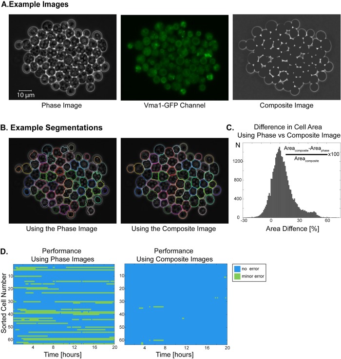
Fig 8. Utilizing a fluorescent channel for improving the segmentation of sporulating cells.
(A) Example phase image, GFP-channel image and the composite image. In the phase image, spores have very bright patches unlike cycling cells. The composite image is created using the phase and GFP-channel images. Note that Vma1-GFP channel is not dedicated to segmentation. (B) Segmentation results using the phase image and using the composite image. Using the composite image corrects for the slight out of focus phase image and significantly improves the segmentation. (C-D) Comparison of segmentations with phase and composite images. Example cells were imaged for 20 hours (100 time points) and segmented with phase or the composite images. (C) Out of 32868 cell segmentation events, 89.5% of them have a greater area when the composite image is used for segmentation. (D) Comparison of errors in segmentation with phase or composite images. Blue no error, green minor error. Minor errors decreased significantly when composite images were used for segmentation.

