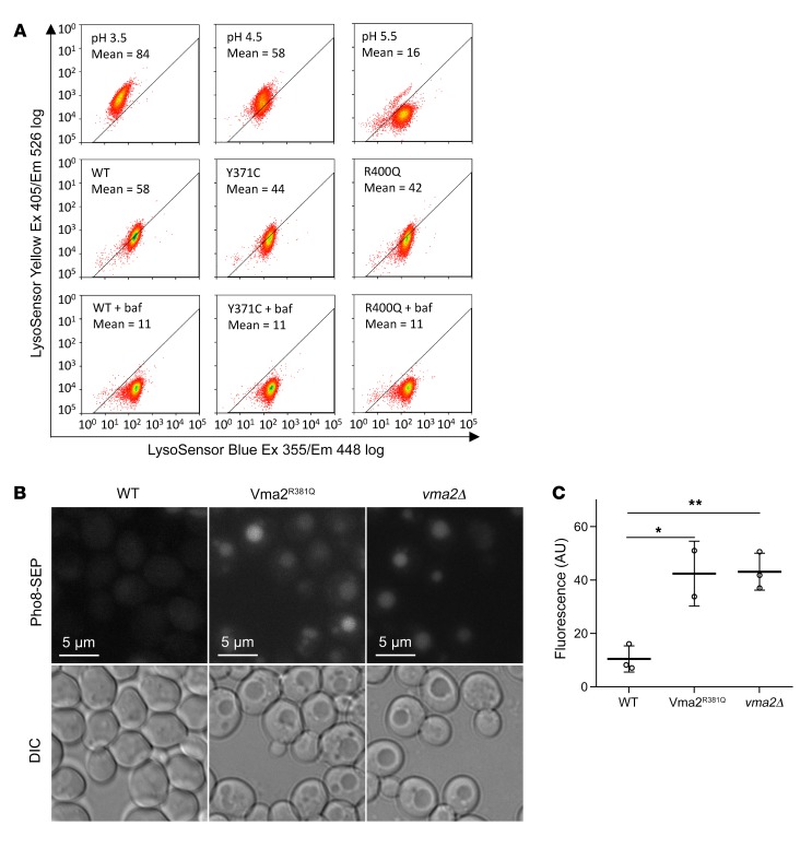
Figure 5. FL-associated ATP6V1B2 mutations reduce the ability of the v-ATPase to acidify lysosomes.
(A) Stable HEK293T cells were generated using the doxycycline-inducible lentivirus pCW57.1 carrying WT or mutated cDNAs encoding ATP6V1B2. Cells were induced with doxycycline for 48 hours and loaded afterwards with a pH indicator dye (dextran-conjugated LysoSensor Blue/Yellow) for 12 hours. In parallel, untransfected HEK293T cells, serving as pH controls, were treated with EMS buffer (see Methods) calibrated to pH 3.5, pH 4.5, or pH 5.5. The fluorescence intensity of cell suspensions was read at various wavelengths using flow cytometry. The MFI of the yellow dye fluorescence signal is a measure of lysosomal pH. Bafilomycin A1 (baf), which completely blocks lysosomal acidification, was used as a control for neutral pH. Ex, excitation; Em, emission. (B) In S. cerevisiae, the C-terminus of the protein Pho8 was tagged with a pH-sensitive SEP protein that increases fluorescence with increasing pH. Like vma2Δ (a knockout strain of the yeast homolog of human ATP6V1B2), Vma2R381Q cells demonstrated increased vacuolar pH compared with WT cells. Upper images show the fluorescence signal; lower images show the corresponding light microscopy. (C) Summary of the fluorescence intensity of vacuolar fluorescence measurements in S. cerevisiae for the data in B. *P < 0.05 and **P < 0.01, by t test with Bonferroni’s correction. Data represent the SD.

