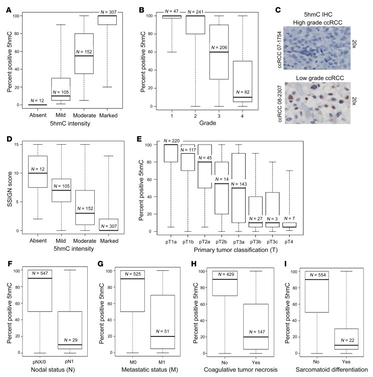
Figure 1. Loss of 5hmC is strongly associated with features of tumor aggressiveness in ccRCC.
(A) Correlation between median percentage positive for 5hmC and 5hmC intensity in IHC (P < 0.001). (B) Higher grade ccRCC is associated with loss of 5hmC (P < 0.001). (C) Representative photographs of low-grade and high-grade ccRCC with 5hmC IHC. (D) Loss of 5hmC correlates with higher SSIGN score, which predicts increased risk of progression of ccRCC after nephrectomy (P < 0.001). (E) Increased tumor size in ccRCC is associated with loss of 5hmC (P < 0.001). (F) Nodal metastasis in ccRCC is associated with loss of 5hmC (P < 0.001). (G) Presence of systemic metastatic disease in ccRCC is associated with loss of 5hmC (P < 0.001). (H) Presence of coagulative tumor necrosis is associated with loss of 5hmC (P < 0.001). (I) Presence of sarcomatoid differentiation is associated with loss of 5hmC (P < 0.001). Box plots have horizontal lines at the 25th percentile, the median, and the 75th percentile. The vertical lines extend to the minimum and maximum values. Associations of 5hmC expression with the clinical and pathologic features studied were evaluated using Spearman’s rank correlation coefficients, Kruskal-Wallis tests, and Wilcoxon’s rank sum tests.

