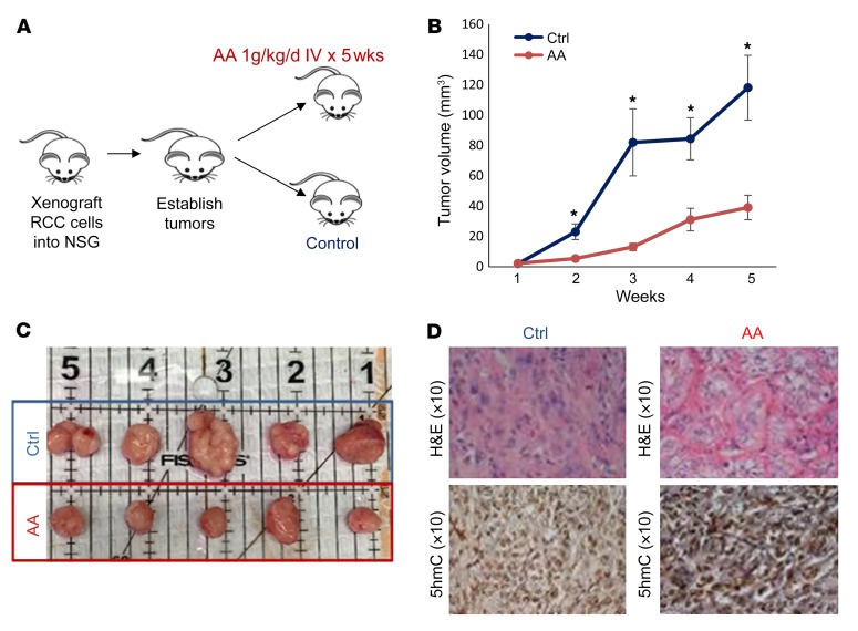
Figure 7. High-dose AA treatment leads to inhibition of ccRCC tumor growth in vivo.
(A) ccRCC cells (786-O) were xenografted into immunodeficient NSG mice. After tumors were established, treatment was initiated with i.v. AA (1 mg/kg/d) or vehicle and tumor measurements were conducted. (B and C) AA treatment led to significantly delayed tumor growth. t test, P < 0.05. Data are shown as mean ± SEM. n = 10 in each cohort. (D) Representative images of histologic examination showing the H&E and 5hmC IHC comparison of ccRCC xenograft control group versus i.v. AA-treated group. Tumor cells in the control group showed a higher grade (poorly differentiated) based on increased prominent hyperchromatic nucleoli, nuclear pleomorphism, multilobation, and multinucleate giant cells when compared with the i.v. AA-treated group. Tumor cells in the i.v. AA-treated group showed a higher intensity and increased staining of nuclei with 5hmC when compared with the control group.

