
Fig. 1. Elastocapillary self-assembly of Neurotassels.
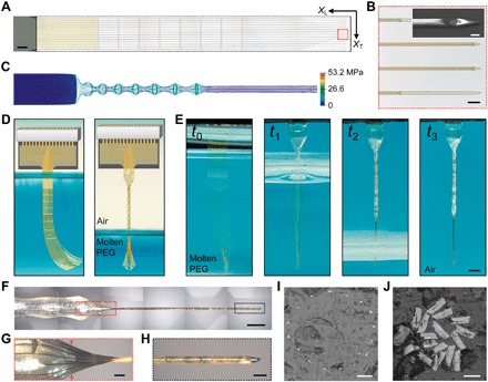
(A) An as-fabricated 16-channel Neurotassel. The black dashed box highlights the freestanding segment supported on an aluminum release layer. XT and XL are the transverse and longitudinal directions, respectively. Scale bar, 500 μm. (B) Zoom-in view of 12-μm-wide and 3-μm-high microelectrode filaments, as marked by the dashed red box in (A). Scale bar, 50 μm. Inset: Scanning electron microscopy (SEM) image of a microelectrode filament with a 10-μm-diameter recording site. Scale bar, 10 μm (inset). (C) Simulated von Mises stresses in a deformed Neurotassel. (D) Schematics of the elastocapillary self-assembly of a Neurotassel. (E) Time sequence photographs of the elastocapillary self-assembly of a Neurotassel. Scale bar, 1 mm. (Photo credit: Shouliang Guan and Jinfen Wang at NCNST). (F) A Neurotassel/PEG assembly. Scale bar, 500 μm. (G and H) Zoom-in views of the Neurotassel/PEG assembly as marked by the red and black dashed boxes, respectively, in (F). Scale bars, 100 μm. (I) Cross-sectional image at the mesh-filament transition section, indicated by the red arrows in (G). Scale bar, 50 μm. (J) Cross-sectional image of the fiber. Scale bar, 10 μm.