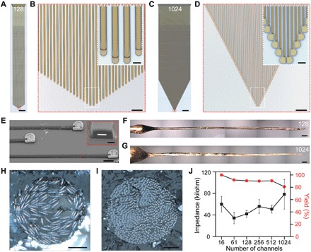
Fig. 6. Scalability of Neurotassels.

(A) An as-fabricated 128-channel Neurotassel. Scale bar, 1 mm. (B) Zoom-in view of the microelectrode filaments in the red dashed box in (A). Scale bar, 50 μm. Inset: Zoom-in view in the white dashed box. Scale bar, 10 μm (inset). (C) An as-fabricated 1024-channel Neurotassel. Scale bar, 1 mm. (D) Zoom-in view of the microelectrode filaments in the red dashed box in (C). Scale bar, 50 μm. Inset: Zoom-in view in the white dashed box. Scale bar, 10 μm (inset). (E) An SEM image (tilted at 45°) of Pt-coated microelectrode filaments of a released 1024-channel Neurotassel. Scale bar, 10 μm. Inset: Focused ion beam–polished cross-section along the red dashed line. Scale bar, 1 μm (inset). (F and G) Assembled 128- and 1024-channel Neurotassel/PEG composite fibers, respectively. Scale bars, 200 μm. (H and I) Cross-sectional images of the assembled 128- and 1024-channel Neurotassel/PEG composite fibers, respectively. Scale bars, 20 μm. (J) Averaged impedance and yield of 16-, 61-, 128-, 256-, 512-, and 1024-channel Neurotassels after Pt electrodeposition. Error bars represent SE (n = 20).
