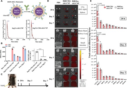
Fig. 4. CD4-targeted LCNPs selectively activate CD4+ T cells from macaque PBMCs and accumulate in the draining LNs after subcutaneous injection to mouse left flank.

(A) Ing3A-LCNP (<150 nm) conjugated with anti-CD4 mAb, resulting in size distribution measured by NanoSight. DSPE, 1,2-distearoyl-sn-glycero-3-phosphoethanolamine; PEG, polyethylene glycol. (B) CD69 median fluorescence intensity (MFI) of CD4+ (CD14−CD3+CD8−) and CD8+ (CD14−CD3+CD8+) cells (left) and their respective MFI ratio (right) from PBMCs isolated from pigtail macaque blood and treated with free Ing3A, bare LCNPs, CD4-targeted LCNPs, and Iso-LCNPs for 20 hours. The experiment was performed once with blood samples from three pigtail macaques (n = 3). (C) Schedule of injection and tissue harvest. C57BL/6J mice were injected with phosphate-buffered saline (PBS), DiR-loaded CD4-cbLCNPs, and Iso-cbLCNPs subcutaneously at the left flank and sacrificed at 20 hours, 3 days, and 7 days for analysis. (D) Representative fluorescent images of inguinal LNs, spleen (top), and other major organs at 20 hours, 3 days, and 7 days. (E) Region of interest quantification of tissue fluorescence normalized by tissue mass. Statistical significance was calculated using paired two-way ANOVA with Bonferroni’s test. **P < 0.005, ***P < 0.005, ****P < 0.0001. Data represent means ± SD; n = 3 mice per group. (Photo credit: Shijie Cao, Department of Bioengineering, University of Washington)
