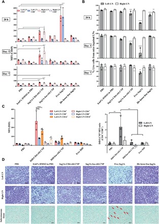
Fig. 5. CD4-targeted LCNPs selectively activate CD4+ T cells in inguinal LNs after subcutaneous injection to mouse left flank and protect local tissues from toxicity.

(A) MFI of CD69 expression on CD4+ (CD14−CD3+CD8−) and CD8+ (CD14−CD3+CD8+) cells from mouse left and right inguinal LNs. Mice were injected with 0.3 ml of PBS, 0.64% DMSO, Ing3A-CD4-cbLCNP (0.48 mg/kg; Ing3A dose per body weight), Ing3A-Iso-cbLCNP (0.48 mg/kg), or free Ing3A (0.48 or 0.024 mg/kg) and were sacrificed after 20 hours, 3 days, and 7 days for analysis. (B) Percentage of live cells from all cell populations in the left or right inguinal LNs after treatment. (C) Left: MFI of DiD fluorescent signal from CD4+ (CD14−CD3+CD8−), CD8+ (CD14−CD3+CD8+), and CD14+ cells from mouse left or right inguinal LNs. Mice were injected subcutaneously at the left flank with 0.3 ml of PBS, DiD/CD4-cbLCNP, DiD/Iso-cbLCNP, or equivalent amount of free DiD (50 μg/kg; DiD dose per body weight) in PBS and were sacrificed after 20 hours for analysis. Right: MFI ratio of DiD fluorescent signal between CD4+ and CD8+ T cells. Statistical significance was calculated using paired two-way ANOVA with Bonferroni’s test comparing each treatment group with PBS. *P < 0.05, **P < 0.005, ***P < 0.0005, ****P < 0.0001. Data represent means ± SD; n = 3 mice per group. (D) Hematoxylin and eosin staining of tissue slides (left and right inguinal LNs and subcutaneous tissues near the injection site) from mice at day 3. From free Ing3A treatment group, condensed cell nucleus in the left LN that indicated cell death and immune cell infiltration into the adipose tissue near the injection site (red arrows) were observed.
