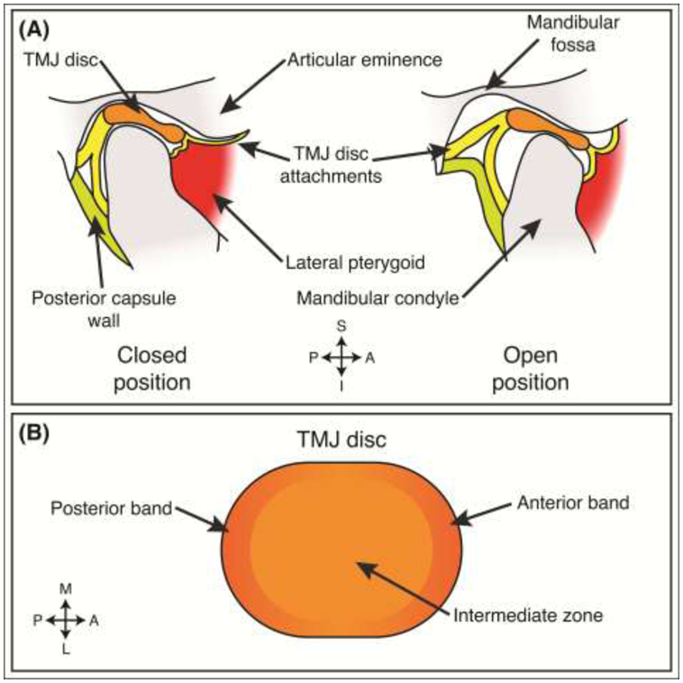
Figure 1: TMJ disc anatomy.

(A) Depending on the open or closed position of the joint, the TMJ disc is situated between the mandibular condyle and the articular eminence-mandibular fossa region. In this sagittal view, the disc is held in place by disc attachments, present at all angles (e.g., lateral, medial, posterior, anterior), surrounding the disc. The joint is separated into two joint capsules delineated by the TMJ disc. (B) The disc is regionally composed of two bands in the anterior and posterior portions of the disc. The middle portion of the disc is referred to as the intermediate zone. S – superior, I – inferior, A – anterior, P – posterior, M – medial, L – lateral.
