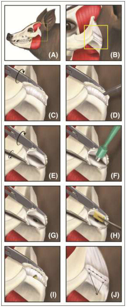
Figure 5: The intralaminar fenestration surgical technique.

(A-B) Through a preauricular incision, the TMJ was exposed. (C-E) Surgeons fileted the disc open with a scalpel, and (F-G) created a one-sided thinning defect via a biopsy punch. (H) A tissue-engineered disc was placed between the two laminae and (I) sutured back together. Sutures attached to the side of the disc instead of on the articulating surfaces allowed for continued loading of the TMJ disc while healing; this placement avoided possible stress concentrations and resulting degeneration. (J) The lateral attachment is recreated by use of an anchoring system. From Vapniarsky, et al., 2018 [39]. Reprinted with permission from AAAS.
