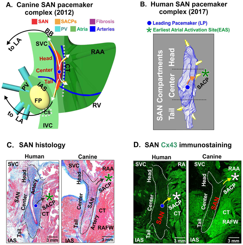
Figure 1: Structural and functional characteristics of the canine and human sinoatrial node (SAN) pacemaker complex.
(A) 3 dimensional model of the canine SAN based on structural and functional data from intramural optical mapping. Compact SAN (red) is isolated from the surrounding atrial myocardium (green) by bifurcating coronary arteries (blue) and fibrotic insulation (purple). Preferential sinoatrial conduction pathways (SACPs) are depicted by yellow bundles and arrows that electrically connect SAN to the atrium. (B) 3-dimensional reconstruction of the human SAN showing intranodal pacemaker compartments and 5 SACPs. (C) Histological features of the canine and human SAN pacemaker complex revealed by Masson’s trichrome staining. (D) Immunostaining identifies the SAN by negative connexin 43 (Cx43) expression. [(A) Data modified from Fedorov et al. Am J Physiol Heart Circ Physiol 2012 [37]; (B) (C) and (D) Figures modified from Li et al. Sci Transl Med 2017 (Human) [11] and Lou et al. Circulation 2014 (Canine) [19]]. BB: Bachmann bundle; CT: crista terminalis; EAS: earliest atrial activation site; FP: fat pad; IAS: interatrial septum; PV: pulmonary veins; RA: right atrium; RAA: right atrial appendage; RAFW: right atrial free wall; SAN: sinoatrial node; SVC: superior vena cava.

