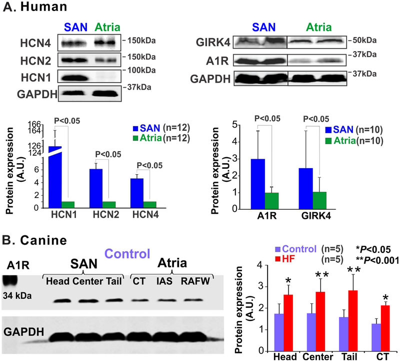
Figure 6: Heterogeneous protein expression in the sinoatrial node (SAN) vs. adjacent atrial tissue.
(A) Protein expression of hyperpolarization-activated cyclic nucleotide-gated channel (HCN) 1, 2 and 4 isoforms (left) and adenosine A1 receptor (A1R) and G protein-coupled inwardly rectifying potassium channel (GIRK4) (right), and summary data below show that these proteins are significantly higher in the SAN relative to atrial tissues; (B) Left, immunoblots showing protein expression pattern of A1R in the SAN compartments and in the atrial myocardium of control canine hearts ; right, summary of these data shows that A1R expression is significantly upregulated in the SAN compartments in heart failure (HF). [(A) Li et al. Circ Arrhythm Electrophysiol. 2015 [58] and Li et al. Sci Transl Med 2017 [11]; (B) Data modified from Lou et al. Circulation 2014 [19] (left)]. CT: crista terminalis; GAPDH: Glyceraldehyde 3-phosphate dehydrogenase; IAS: interatrial septum; RAFW: right atrial free wall.

