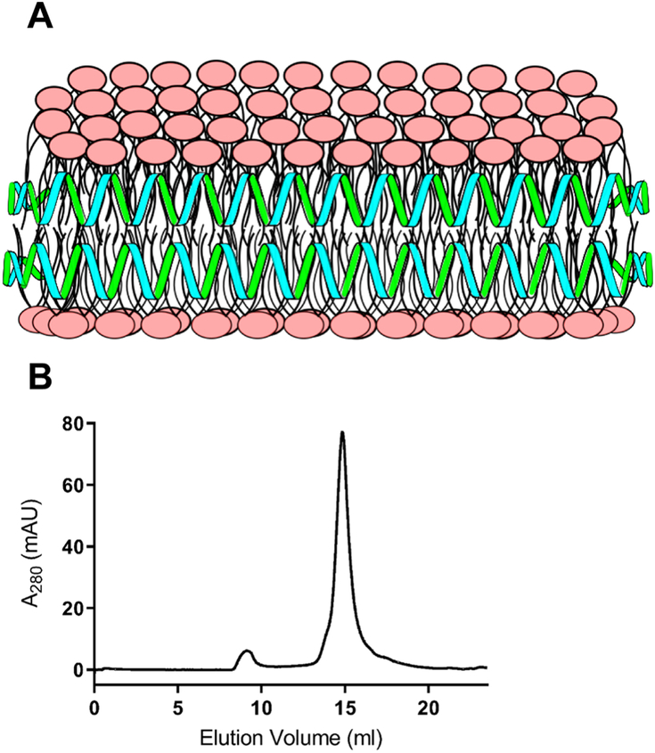
Figure 1: Model structure of CL ND.
A) In this depiction, CL (polar head groups in pink) is organized as a disk-shaped bilayer whose perimeter is circumscribed and stabilized by apoA-I (green/blue helices), which interacts with CL fatty acyl chains at the edge of the disk. B) FPLC size exclusion chromatography profile of CL ND. ND were formulated with apoA-I and CL as described. An aliquot of CL ND (0.32 mg apoA-I / 0.8 mg CL) in 20 mM HEPES buffer, pH 7.2, was applied to a Superose 6 Increase 10/300 GL column and elution monitored at 280 nm.

