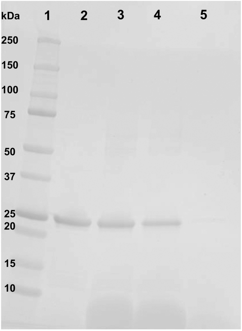
Figure 6. Effect of CaCl2-mediated disruption of CL ND on apoA-I solubility.
CL ND were formulated with apoA-I in HEPES buffer and an aliquot, corresponding to 78 nmol CL, was incubated with 2000 nmol CaCl2 for 1 h at 22 °C to induce sample turbidity development. Following this, the sample was centrifuged at 14,000 × g for 2 min and the supernatant recovered. The precipitate was re-suspended in a volume of HEPES buffer equal to that of the reserved supernatant. Equivalent aliquots of the resuspended pellet and supernatant were electrophoresed on a 4–20% acrylamide gradient SDS PAGE gel and stained with GelCode Blue. Lane 1) molecular weight markers; Lane 2) apoA-I standard; Lane 3) control CL ND; Lane 4) CaCl2-disrupted CL ND re-suspended pellet; Lane 5) CaCl2-disrupted CL ND supernatant.

