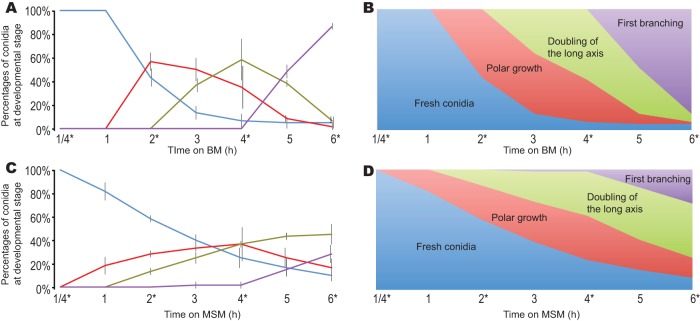
FIG 2.
Temporal analysis of growth and development of conidia of N. crassa cultured on BM and MSM. Plated conidia were examined at six time points across the process, enabling sector counts that revealed the (A) proportions and (B) stacked proportions of conidia at serial stages of germination cultured on BM and the (C) proportions and (D) stacked proportions of conidia at serial stages of germination cultured on MSM. Measurements for conidia were color-coded at each stage of germination, including those corresponding to fresh conidia (blue), polar growth (red), doubling of the longest axis (green), and first hyphal branching (purple). An asterisk (*) indicates time points when RNAs were sampled. For staging, germination of 20 randomly selected conidia per plate was monitored; the error bars delineate 1 standard deviation of the mean for three such plates.

