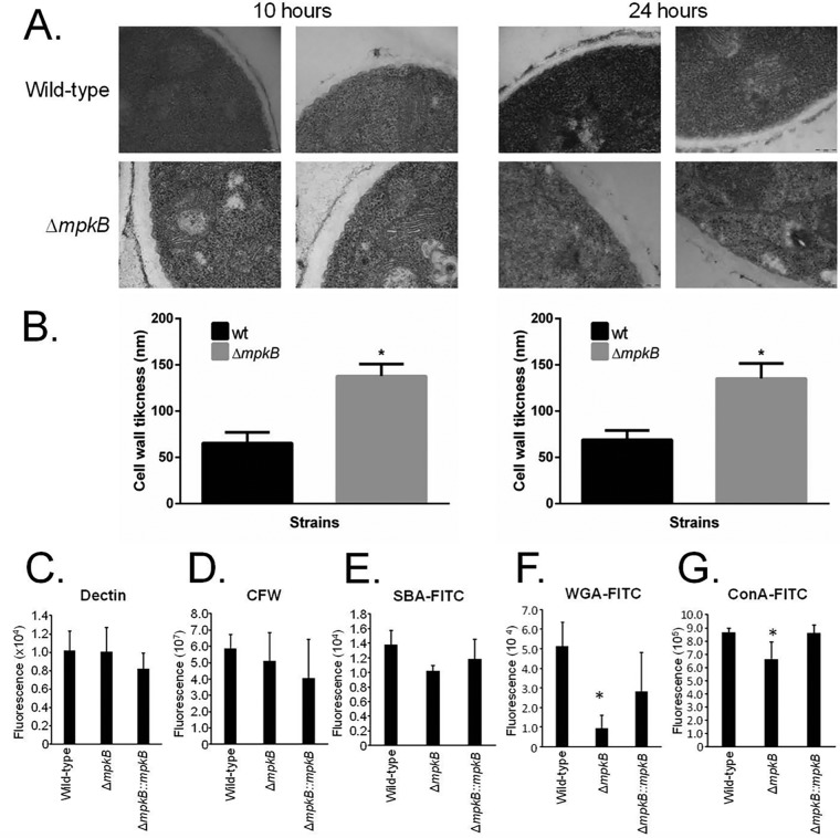
FIG 5.
The ΔmpkB mutant strain has an altered cell wall organization. (A) Transmission electron microscopy of mycelial sections of the A. fumigatus wild-type and ΔmpkB strains grown for 10 h or 24 h in MM at 37°C. (B) The cell wall thicknesses (nm) of 100 sections of different hyphal germlings (average of 4 sections per germling) were measured when grown under the same conditions as specified in the legend to panel A. The averages and standard deviations of the 50 measurements are presented. Statistical analysis was performed using the one-tailed, paired t test, comparing data to the results for the control condition (*, P < 0.00001). (C to G) Detection of different sugars exposed on the cell surface. Conidia were cultured in liquid MM to the hyphal stage, UV killed, and stained with CFW, soluble dectin-1, or specific probe to detect the content of exposed sugars. Experiments were performed in triplicate, and the results are displayed as mean values with standard errors (two-way ANOVA followed by Tukey’s; *, P < 0.05).

