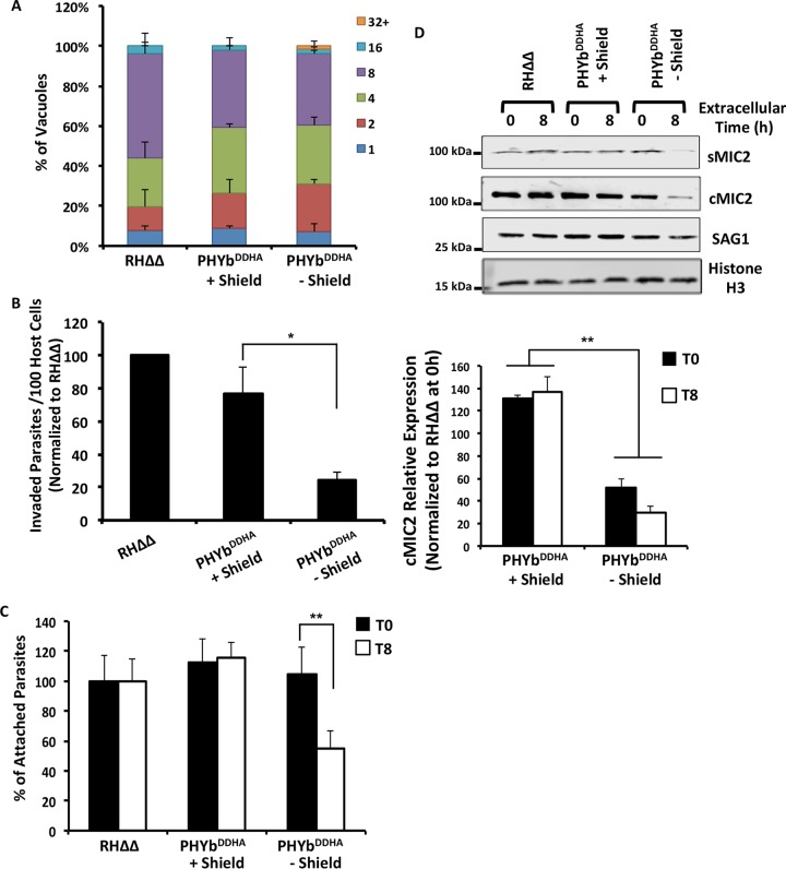
FIG 5.
Extracellular stress leads to reduced host cell invasion by TgPHYb-depleted parasites. TgPHYbDDHA parasites grown for 24 h with or without Shield-1 were harvested and exposed to stress for 0 or 8 h at 21% O2. (A) Replication of extracellularly stressed parasites was determined by counting numbers of parasites per vacuole 24 h postinfection. At least 100 vacuoles per strain were counted, and shown are the averages and standard deviations from three independent assays. (B) Parasites were added to HFF monolayers in high-K+ buffer for 20 min and replaced with prewarmed invasion medium for 1 h. The cells were fixed, and numbers of intracellular parasites were determined by differential SAG1 staining and normalized to RHΔΔ parasites. *, P < 0.05, Student’s t test. (C) Parasites were added to HFF monolayers in high-K+ buffer for 20 min. The cells were then washed to dislodge weakly associated parasites, fixed, and stained with SAG1 antisera to determine numbers of intimately attached parasites at each time point, normalized to parental RHΔΔ strain parasites. **, P < 0.005, Student’s t test. (D) Parasites were incubated with 1% (vol/vol) ethanol for 5 min to stimulate microneme discharge. After centrifugation, supernatants and parasites (from equal cell numbers) were separated by SDS-PAGE and Western blotted to detect secreted MIC2 (sMIC2), cell-associated MIC2 (cMIC2), SAG1, and Toxoplasma histone H3 as a loading control. Shown are the average and standard deviations from 3 independent experiments as well as representative blots.

