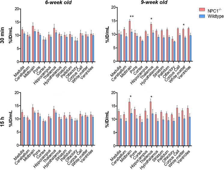
Fig. 6.
Top: Regional brain distribution of 64Cu in 6-week-old (left panels) and 9-week-old (right panels) Npc1−/− mice and age-matched controls at 30 min (top panels) and 15 h (bottom panels) post-injection of 64Cu-GTSM. Data are mean [n = 6-7 Npc1−/−, n = 5 WT)] ± SD. Statistically significant differences from healthy controls were evident in the 9-week-old groups and are indicated by * (p < 0.05) and ** (p < 0.01)

