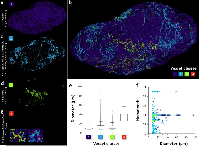
Figure 6.
Intra-tumor heterogeneity results in the establishment of unique microvascular ‘niches’. Microvasculature in Day 21 tumor (Tumor 1) is color coded into four classes based on their physiological characteristics and vascular structure. Class 1 vessels (purple) (a) were hypoperfused and hypoxic (i.e. Vel < 50 µm/s and PO2 < 10 mmHg); Class 2 vessels (light blue) (c) were hyperperfused and normoxic (i.e. Vel ≥ 50 µm/s and PO2 ≥ 10 mmHg); Class 3 vessels (light green) (d) were either hypoperfused and normoxic or hyperperfused and hypoxic, and finally Class 4 vessels (red) (g) served as functional ‘shunts’ (i.e. velocity at least 2× larger than the median blood flow velocity, vessel diameter at least 2× greater than the median diameter, and vascular length smaller than the median length by a factor of 0.5). Since occurrence of Class 4 vessels is infrequent, the inset shows zoomed images illustrating their role as functional shunts within the tumor. (b) Spatial arrangement of these vessel classes in 3D. (e) Box and whisker plots of diameter (μm) for each vessel class, (f) plot of hematocrit vs. diameter (μm) for plasma-bearing vessels in each class.

