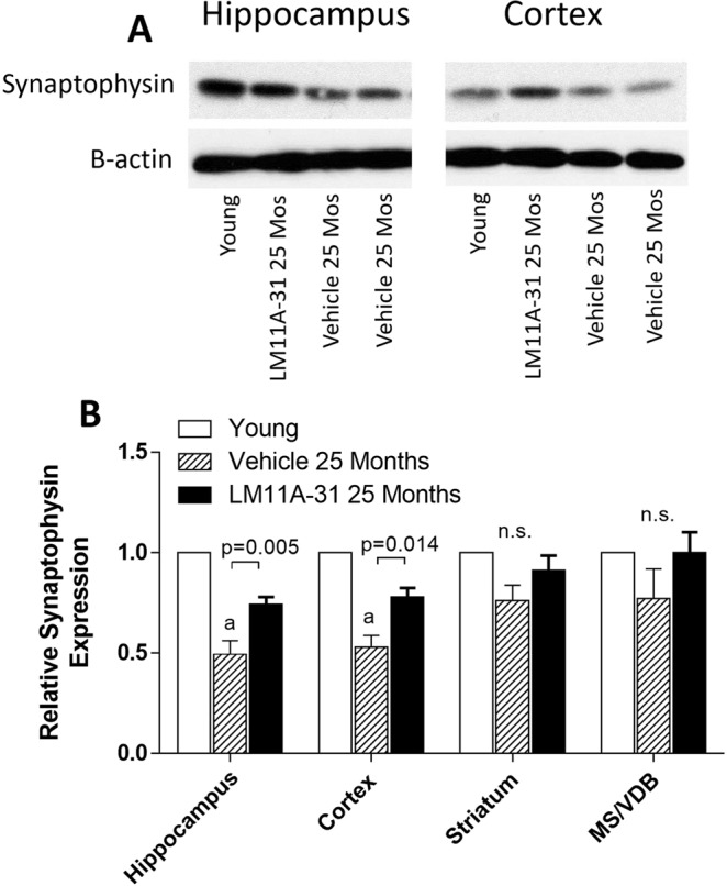
Figure 9.

Western blot analysis of synaptophysin levels in the hippocampus of 4-month-old mice and 25-month-old mice treated with 50 mg/kg LM11A-31 or vehicle (saline) from 21 to 25 months. (A) In the hippocampus and cortex, synaptophysin was reduced in aged mice relative to young mice and partially restored by treatment with LM11A-31. The bands shown are representative of the differences seen across nine different blots and were acquired from the gel scans in Supplemental Fig. S1. Beta actin bands were generated from the same blot after stripping and re-probing. (B) Synaptophysin signal normalized to beta-actin showed significant age-related reduction in hippocampus, cortex and striatum with a significant recovery of signal associated with LM11A-31 treatment in the hippocampus and cortex (p’s = 0.005–0.014, t test, treated vs vehicle, n = 9). Values are mean + sem. a, P < 0.001 relative to young mice.
