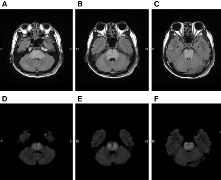
Fig. 2.
a–c T2-weighted fluid-attenuated inversion recovery magnetic resonance imaging (MRI) images. d–f Diffusion-weighted magnetic resonance imaging shows multifocal abnormal signal intensity change in the whole brain stem; these lesions involve the parts from the lower midbrain and whole pons to the upper medulla oblonganta

