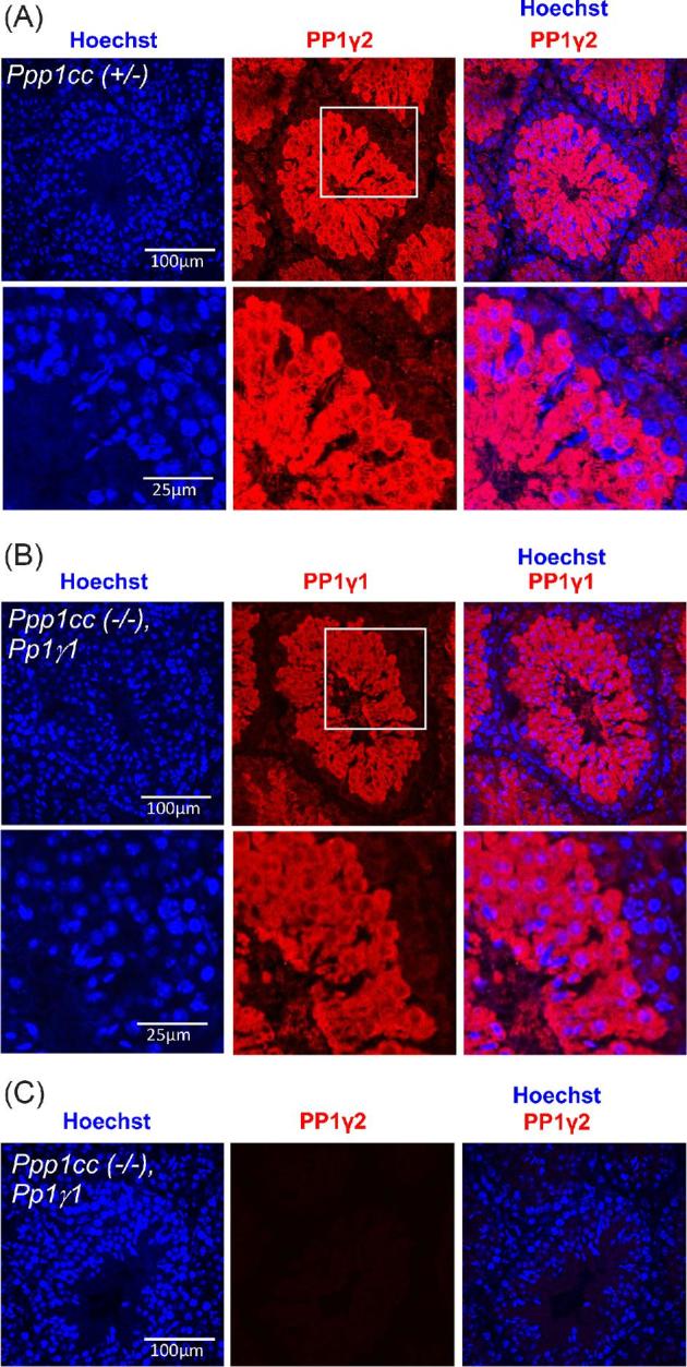
Figure 4.

Immunohistochemisrty showing the expression and localization of PP1γ2 in Ppp1cc (+/–) and PP1γ1 in Ppp1cc (–/–), Pp1γ1 testis. (A) Ppp1cc (+/–) testis sections stained with anti-PP1γ2 antibody (red) and counterstained with Hoechst for nuclei (blue). PP1γ2 staining appears in meiotic and postmeiotic germ cells but absent from premeiotic spermatogonial cells. The region highlighted with white box is enlarged and shown in the bottom panel. (B) Ppp1cc (–/–), Pp1γ1 testis sections stained with anti-PP1γ1 antibody (red) and counterstained with Hoechst for nuclei (blue). PP1γ1 staining appears in meiotic and postmeiotic germ cells but absent from premeiotic germ cells near the basal membrane similar to the expression pattern of PP1γ2 in Ppp1cc (+/–) control testis. The region highlighted with white box is enlarged and shown in the bottom panel. (C) Ppp1cc (–/–), Pp1γ1 testis sections stained with anti-PP1γ2 antibody show no detectable signal (red) indicating the absence of PP1γ2.
