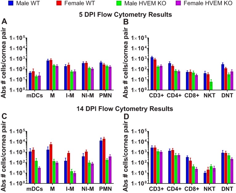
FIG 2.

Immune cells infiltrate male and female WT and HVEM KO corneas at similar levels during the acute (5-dpi) and chronic (14-dpi) phases of HSV-1 corneal infection. Data represent results of flow cytometry analysis of immune cell infiltrates in WT and HVEM KO corneas collected at 5 dpi (n = 8 to 10, two replicates) (A and B) and 14 dpi (C and D). (A and C) Absolute (Abs) number of myeloid infiltrates of dendritic cells (mDCs), macrophages (M), inflammatory macrophages (I-M), noninflammatory macrophages (NI-M), and polymorphonuclear leukocytes (PMN) at 5 dpi (A) and at 14 dpi (C). (B and D) Absolute number of lymphoid infiltrates of CD4+ T cells, CD8+ T cells, NK T cells, or DN T cells per cornea pair at 5 dpi (B) and at 14 dpi (D) (n = 8 to 20 mice, 2 or 3 replicates). No NKT cells were detected in female HVEM KO corneal samples at 5 dpi. There was no significant difference between males and females of either genotype. The differences between the WT and HVEM KO data are consistent with our previous studies. Infiltrating cell percentages (calculated as means ± SEM) were analyzed using two-tailed t tests with Holm-Sidak's correction for multiple comparisons.
