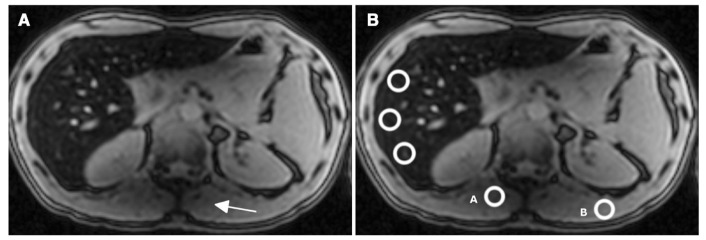
Fig. 6.
This MRI corresponds to the forty-year old male patient marked by an asterisk in Figure 3 (B-HIC = 405 μmol/g and MR-HIC=236μmol/g). First echo (TE=1.15 msec) image. (a) The signal decreases in the left lobe of the liver and in the paraspinal muscles (arrowhead) due to a B1 heterogeneity artifact. (b) The MR-HIC estimation can vary between 190 and 390 μmol/g just by moving ROI A, in the artifact area, or B, outside the artifact area, to determine the muscle signal intensity reference.

