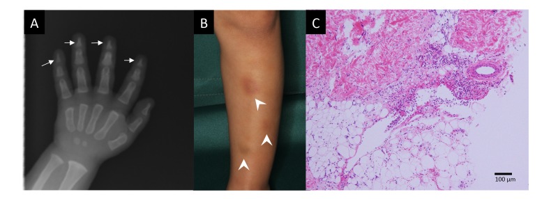
Fig. 1.
A: Skeletal X-ray at 1 year of age showing tapered distal phalanges (arrows). B: Erythema appearing on the anterior surface of the lower leg at 3 years of age (arrow heads). C: Histopathology of an erythema showing perivascular and septal inflammatory infiltration in the subcutaneous adipose tissue (Hematoxylin–Eosin staining, bar = 100 μm).

