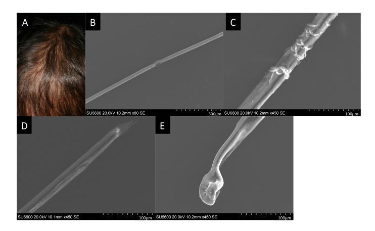
Fig. 2.
A: Sparse scalp hair at 5 years of age. The surface of the scalp is partially exposed because of hair loss. B–E: Scanning electron microscopy images of the patient’s hair at 5 years of age. (B) Trichorrhexis represented as a focal longitudinal vulnerable lesion of the hair shaft. (C) The hair shaft showing a sagged or peeled surface. (D) Trichoptilosis represented as a longitudinal splitting and cracking lesion at the distal end of the hair shaft. (E) Misshapen and depressed hair bulb.

