Figure 1.

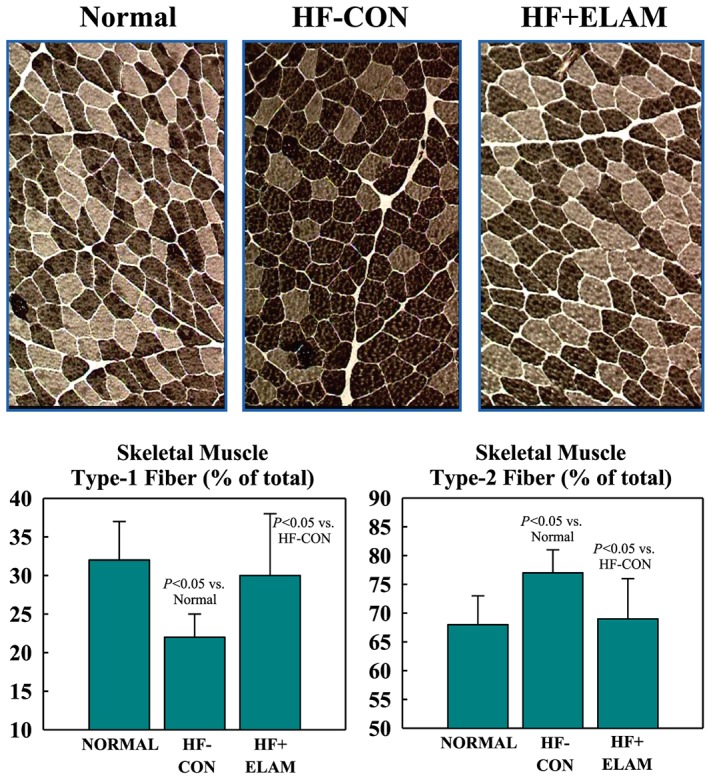
Top: ATPase‐stained histological panels (same magnification) depict skeletal muscle fibre‐type composition in triceps muscle. Type 1 fibres appear as lightly stained fibres (light brown) and type 2 fibres as dark‐stained fibres (dark brown). Left top panel: fibre‐type composition in a normal dog. Top middle panel: fibre‐type composition in an untreated heart failure control (HF‐CON) dog. Note that the number of type 1 fibres is reduced relative to normal dogs in the left panel. Top right panel: fibre‐type composition in an elamipretide‐treated heart failure (HF + ELAM) dog. Note that the number of type 1 fibres is increased relative to HF‐CON in middle panel. The bottom 2 graphs show the numerical distribution (mean ± standard error of the mean) of skeletal muscle type 1 and type 2 fibres in normal dogs and in both study groups.
