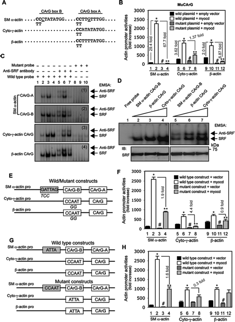
Figure 6.

Myocardin-induced differential regulation of actin isoforms is CArG box and ATTA, CCAAT box dependent. (A) CArG box sequences from rat SM α-actin, cytoplasmic-γ-actin (Cyto-γ-actin) and β-actin promoters are aligned. Site mutations are shown in bold letters (CC in CArG boxes were replaced by TT). (B) Activated HSCs were cotransfected with luciferase reporter constructs containing wild type or the CArG box mutation as in (A) and a myocardin expression plasmid (Myocd) or empty vector. Cells were harvested 2 days later to detect for promoter activity. (C) After growth for 2 days after isolation, HSCs were exposed to the indicated adenoviral vectors for 3 days and 2 days later, they were subjected to EMSA to measure the effect of myocardin on SRF binding activity. SRF binding and supershifted bands are highlighted by arrows (lane 1, 2, 5, 8: nuclear extracts from HSCs infected with Ad-control virus; lane 3, 6, 9: nuclear extracts from HSCs infected with Ad-myocd virus; lane 4, 7, 10: nuclear extracts from HSCs infected with Ad-myocd-DN virus). Representative data from 3 independent experiments are shown. (D) EMSA was performed using nuclear extracts (10 μg) from activated HSCs and the same amount of different actin CArG box probes (1 × 10⁵ cpm) as indicated. SRF binding and supershifted complexes were indicated by arrows (upper panel). Nuclear extracts were probed by anti-SRF antibody as loading control (bottom panel). Representative data from 3 independent experiments are shown. (E) A schematic diagram of wild type and mutant actin isoform promoters is shown (mutated nucleotides were indicated below the consensus sequences); (F) luciferase assays were performed as in (B). (G) A schematic diagram of exchanged elements (ATTA and CCAAT boxes) among SM α-actin, cytoplasmic-γ-actin (cyto-γ-actin) and β-actin promoters is shown; (H) luciferase activity assay was performed as in (B). (n=3, * p < 0.01 for wild plasmid + empty vector vs. wild plasmid + myocardin; n=3, # p < 0.05 for wild plasmid + empty vector vs. mutant plasmid + empty vector; n=3, ** p < 0.05 for wild plasmid + myocardin vs. mutant plasmid + myocardin).
