Figure 1.
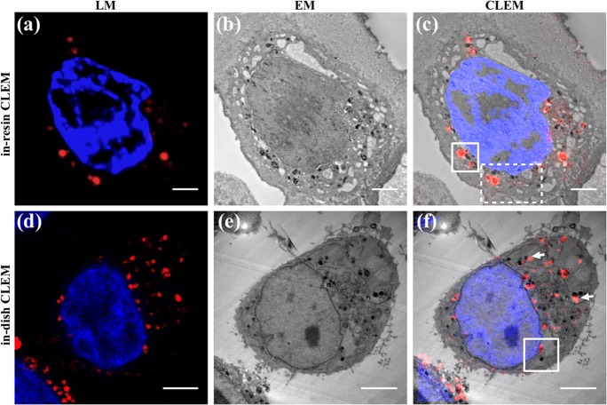
Correlative light-electron microscopy (CLEM) of fNDs in HeLa cells showing the results of the in-resin (top) and in-dish (bottom) preparation. (a) Confocal laser scanning microscopy (CLSM) of fNDs (red) and nucleus (blue, Hoechst) on ultrathin section (120 nm nominal thickness). (b) Transmission electron microscopy (TEM) of the same section as in (a). (c) Overlay of (a,b). (d) A selected image (of LM stack) with fNDs (red) and nucleus (blue, Hoechst) of a paraformaldehyde (PFA) fixed HeLa cell. (e) Corresponding epoxy resin section of the same cell as shown in (d) and the resulting CLEM overlay (f). Scale bar: (a–c) 2 μm, (d–f) 5 μm. The white boxes denoted in (c) refer to the areas displayed in Figure 3, and the box denoted in (f) refers to the area shown in Figure 2.

