Figure 2.
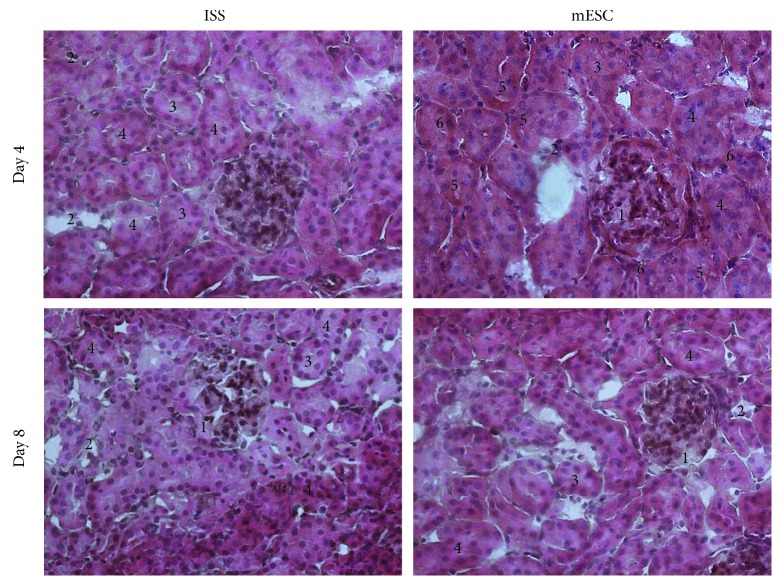
Histopathological analysis. The kidney sections were stained with hematoxylin and eosin, and representative images are shown. The isotonic salt solution (ISS) and the mouse embryonic stem cells (mESC) groups exhibited a segmental focal glomerulosclerosis (1), inflammatory infiltrated (2), tubular dilatation (3), and diffuse denudation of the tubular cells plugging the tubular lumen (4). The mESC group showed tubules less dilated, cytoplasmic vacuoles in the proximal tubular cells (5) and binucleation (6) (n = 5 with three biological replicates, 200x).

