
Fig 3.
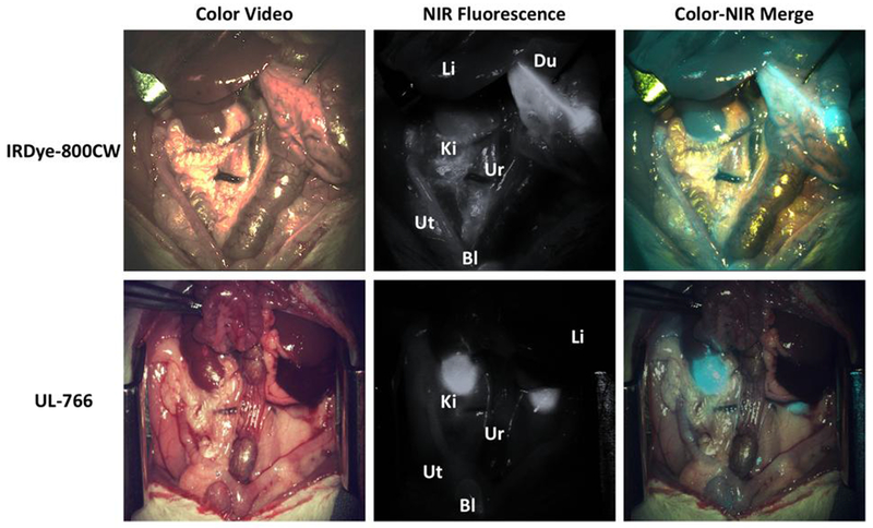
NIR fluorescence-guided intraoperative identification of the ureter. Shown are the color video (left column), NIR fluorescence (middle column), and a pseudo-colored (Cyan) merged image of the 2 (right column). Exposure time was 33 ms for all NIR fluorescence images. Li:liver, Du:Duodenum, Ki:kidney, Ur: ureter, Ut: uterine, Bl: Bladder.