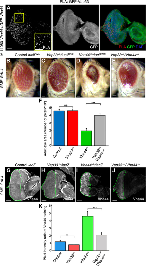
Fig. 6. Vap33 physically and genetically interacts with the V-ATPase component Vha44 and reduces Vha44 protein abundance.

(A) Confocal planar images of the PLA in MI11080 Vha44-eGFP-Vha44 third-instar larval eye-antennal discs using GFP and Vap33 antibodies. The eye-antennal discs were also stained to show Vha44-GFP. Nuclei were stained with DAPI (blue). (B to E) Images of adult female eyes in which the indicated RNA interference (RNAi) or overexpression (o/e) constructs were expressed under the control of the eye-specific GMR-GAL4 driver. (B) GMR>luciferaseRNAi (control). (C) GMR>Vap33o/e and luciferaseRNAi. (D) GMR>Vha44 and luciferaseRNAi. (E) GMR>Vha44 and Vap33. (F) Quantification of eye size (pixels) from experiments illustrated in (B) to (E). n = 3 independent experiments, n = 3 samples per genotype per experiment. Error bars indicate SEM. ***P < 0.0001; ns, differences not significant (t tests with two-tailed distribution and unequal or equal variance). (G to J) Confocal planar images of third-instar larval eye-antennal discs from flies expressing the indicated transgenes under the control of the GMR-GAL4 driver and stained to show Vha44. (G) GMR>lacZ (control). (H) GMR>Vap33 and lacZ. (I) GMR>Vha44 and lacZ. (J) GMR>Vha44 and Vap33. Scale bars, 50 µm. (K) Quantification of the pixel intensity ratio of the mutant GMR posterior domain compared to the anterior WT domain from eye discs in (G) to (J). n = 3 independent experiments, n = 3 samples per genotype per experiment. Error bars indicate SEM. ***P < 0.0001, *P = 0.008. ns, differences not significant (t tests with two-tailed distribution and unequal or equal variance).
