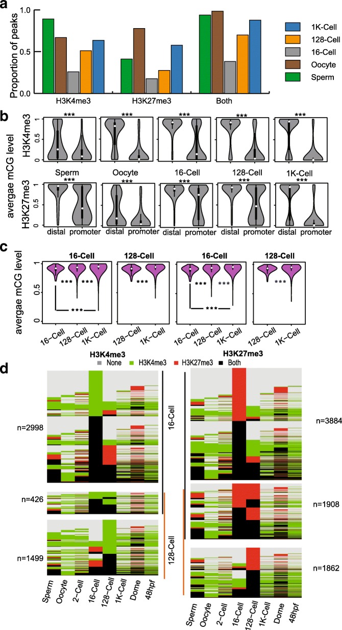
Fig. 3.
DNA methylation patterns in distal and promoter peaks. a Bar plot for proportion of peaks that overlap with hypomethylated regions in each stage. b Violin plot demonstrates the differences between distal and promoter peaks in each stage for H3K4me3 and H3K27me3. c Violin plot showing the DNA methylation pattern dynamics in hypermethylated promoter peaks found in 16-cell and 128-cell stages for H3K4me3 and H3K27me3. d Histone modification dynamics for hypermethylated promoter peak-associated genes found in the 16-cell and 128-cell stages. Note: ‘***’ denotes that there is a significant difference detected by the Wilcox test

