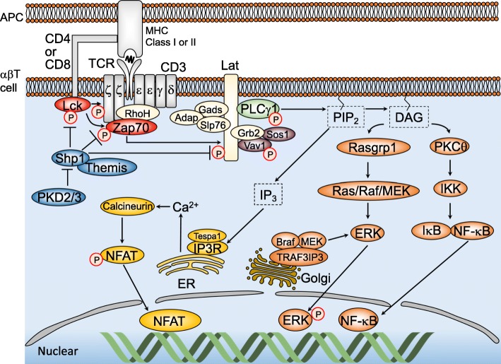
Fig. 1.
Schematic diagram of αβT cell receptor (TCR) signaling pathway. TCR engagement by pMHC expressed on antigen-presenting cells (APC) induces phosphorylation of CD3 ITAMs by Lck. Zap70 binds to phosphorylated ITAMs and is phosphorylated as well by Lck. The activated Zap70 then phosphorylates Lat, which induces recruitment of adaptor proteins (Gads, Adap, Slp76, and Grb2) and signaling molecules (PLCγ1, Sos1, Vav1). Phospho-PLCγ1 catalyzes hydrolysis of PIP2, resulting in generation of DAG and IP3. DAG leads to translocation of Rasgrp1 and PKCθ to the plasma membrane, resulting in activation of Ras/MAPK pathway and NF-κB pathway. IP3 stimulates endoplasmic reticulum (ER) for the releases of calcium ions, which activate NFAT pathway. Shp1 dephosphorylates a broad range of signaling molecules including Lck, CD3, Zap70, Lat, Slp76, and Vav1, to finely tune TCR signal

