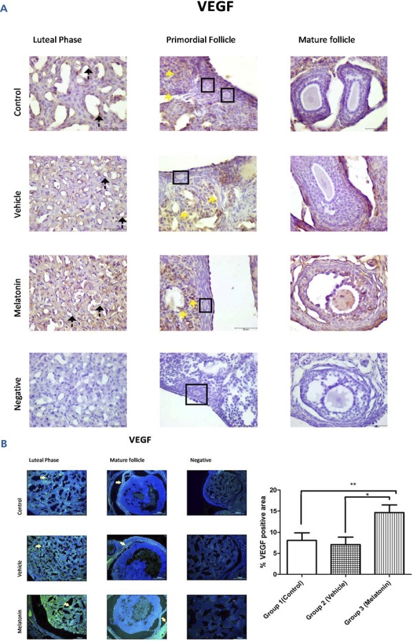
Figure 3. Immunoreactivity of VEGF proteins.

A: Immunohistochemical staining, B: Immunofluorescence staining
Immunoreactivity of VEGF proteins in Group 1 (control), Group 2 (vehicle), and Group 3 (melatonin). An increase in VEGF in the endothelial cells of the blood vessel (as indicated by yellow arrows) can be seen in the melatonin treatment group. Data are displayed as the mean ± S.E.M.; n=15 rats/group. **, p <0.05 indicates significance compared to the respective control values. *, p <0.05 indicates significance compared to the respective vehicle values.
