Figure 5.
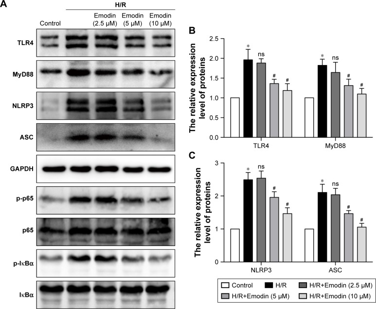
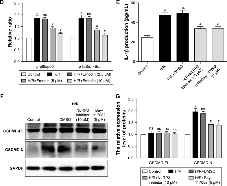
Inhibition of pyroptosis by emodin involved the TLR4/MyD88/NF-κB/NLRP3 inflammasome pathway.
Notes: (A) Representative Western blot luminogram of TLR4, MyD88, NLRP3, ASC, p-p65, and p-IκBα among the control, H/R, H/R+Emodin (2.5 µM), H/R+Emodin (5 µM), and H/R+Emodin (10 µM) groups. Cells in normoxic conditions were defined as the control (B). Protein semiquantification is shown for TLR4 and MyD88 based on the results of 5A. (C) Protein semiquantification is shown for NLRP3 and ASC based on the results of 5A. (D) Protein semiquantification is shown for p-p65 and p-IκBα based on the results of 5A. (E) Concentrations of IL-1β in cell culture supernatants were detected by ELISA. (F) Representative Western blot luminogram of GSDMD-FL and GSDMD-N among the control, H/R, H/R+DMSO, H/R+NLRP3 inflammasome inhibitor (10 µM), and H/R+Bay-117082 (5 µM) groups. (G) Protein semiquantification is shown for GSDMD-FL and GSDMD-N based on the results of 5F. Bay-117082, NF-κB pathway inhibitor. p-p65 was standardized by p65, and p-IκBα was standardized by IκBα. The expression of other proteins was standardized by GAPDH. Data are expressed as the mean ± SD. *P<0.05 vs the control group, #P<0.05 vs the H/R group.
Abbreviations: H/R, hypoxia/reoxygenation; DMSO, dimethyl sulfoxide; ns, not significant.


