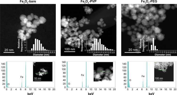
Figure 1.
Characterization of IONP by TEM.
Notes: Micrographs of three different IONP and their size distribution (upper panel). Elemental mapping by energy-dispersive X-ray spectroscopy (lower panel). Abbreviations: IONP, iron oxide nanoparticles; PEG, polyethyleneglycol; PVP, polyvinylpyrrolidone; TEM, transmission electron microscope.

