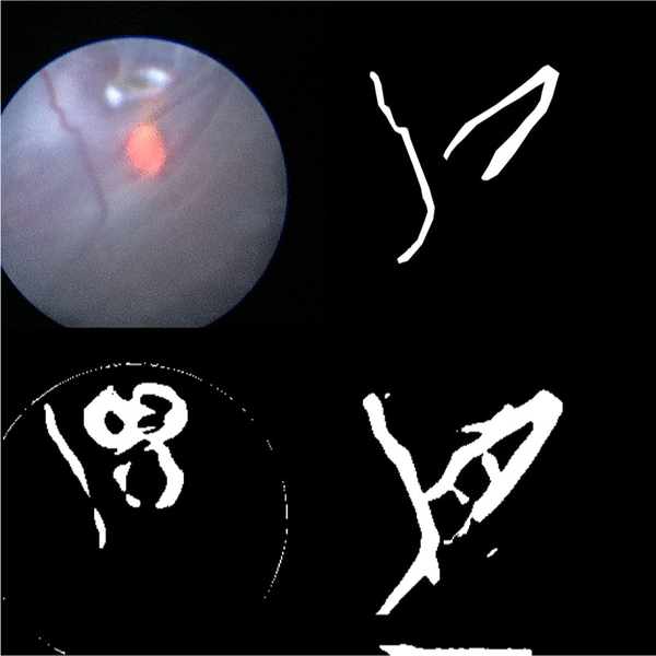
Fig. 4.

The results of running various segmentation algorithms on a particularly difficult fetoscopic video frame that was not included in the training data. Top left: the input fetoscopic video frame. Top right: the ground truth segmentation. Bottom left: the segmentation generated by thresholding the Frangi filtered image. The Frangi filter mistakenly identifies the glare at the top of the image and the guide light at the center of the image as blood vessels. Bottom right: the segmentation generated by the FCNN. Note the close correspondence to the image created by the human rater
