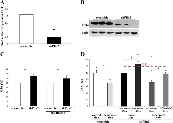
Figure 1.

Plin2 down‐regulation in adult tibialis anterior (TA) muscle fibres. (A and B) RNAi‐mediated knockdown of Plin2 revealed by real‐time RT–PCR (* P < 0.001) (A) and immunoblotting (B). (C and D) Cross‐sectional area (CSA) of TA muscles upon knockdown of Plin2 expression. (C) CSA of adult TA muscle fibres transfected with either shPlin2 or scramble. Left columns: * P = 0.001; right columns: the same, plus treatment with rapamycin: * P = 0.02. (D) CSA of adult TA muscle fibres transfected with shPlin2 or scramble and undergone denervation performed at the same time of transfection. Left columns: CSA of TA muscle fibres transfected with scramble; control vs. denervated, * P = 0.001. Right columns: CSA of TA muscle fibres transfected with shPlin2; NO‐shPlin2 indicates fibres that remained untransfected; GFP‐shPlin2 indicates fibres transfected with the shPlin2; * P < 0.001. In all cases, transfected fibres from six mice for each group were identified by GFP fluorescence, and a minimum of 700 fibres for each muscles were measured. Data are shown as mean ± SEM. P values refer to two‐tailed Student's t‐test.
