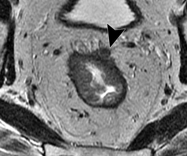
Figure 10d.
Rectal MR images that show distinct tumor stages obtained from three different patients. (a, b) Sagittal (a) and axial (b) T2-weighted MR images show a polypoid lesion (solid arrow) surrounded by mucoid material, with a thin stalk attached to the rectal wall and the intact muscularis propria (dashed arrow), findings characteristic of a T1 or T2 tumor. (c) Oblique axial T2-weighted MR image in another patient shows a tumor infiltrating 7 mm beyond the muscularis propria (T3c), with positive MRF infiltration (arrowhead). (d) Oblique axial T2-weighted MR image in a third patient shows a tumor invading the anterior peritoneal reflection (arrowhead), a characteristic finding of a T4a grade tumor.

