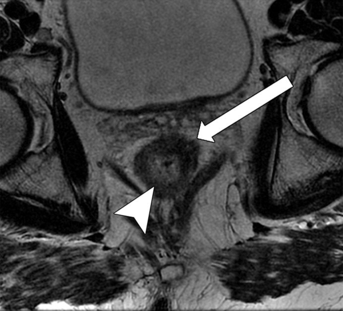
Figure 14c.
Low rectal cancer in a 56-year-old man who underwent neoadjuvant CRT and had clinical complete response, with tumor regrowth 9 months later. (a, b) Oblique axial T2-weighted MR image (a) obtained during primary staging shows an infiltrative tumor (arrow) with the most invasive border between the 1-o’clock and 3-o’clock positions, infiltrating 1 mm beyond the muscularis propria (T3a), which corresponds to a polypoid lesion seen on the colonoscopic image (b). (c, d) Oblique axial T2-weighted MR image (c) obtained after neoadjuvant CRT shows an area with low signal intensity (arrow), and colonoscopic image (d) shows a scar in the tumor bed, without residual tumor. Note the wall thickening and mucosal edema within the normal rectal wall, which were caused by CRT (arrowhead in c). The patient was selected for a watch-and-wait protocol. (e) Oblique axial T2-weighted MR image obtained 9 months later shows thickening in the tumor bed, with areas of intermediate signal intensity (arrow), a finding suspicious for tumor regrowth. (f) Colonoscopic image shows tumor regrowth.

