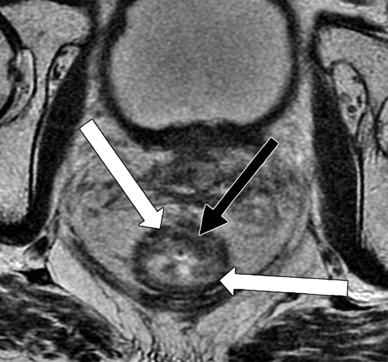
Figure 15c.

Partial response after neoadjuvant CRT. (a, b) Oblique axial T2-weighted MR image (a) and diffusion-weighted image (b) obtained during primary staging shows a tumor (arrow) in the lower rectum, with intermediate signal intensity in a and restricted diffusion in b. (c) Oblique axial T2-weighted MR image obtained after neoadjuvant CRT at restaging shows areas of low signal intensity (black arrow) in the tumor bed and residual tumor (white arrows) with intermediate signal intensity. (d, e) Axial diffusion-weighted image (d) shows restricted diffusion within the areas of the residual tumor (arrowhead), which was confirmed on the corresponding ADC map (e).
