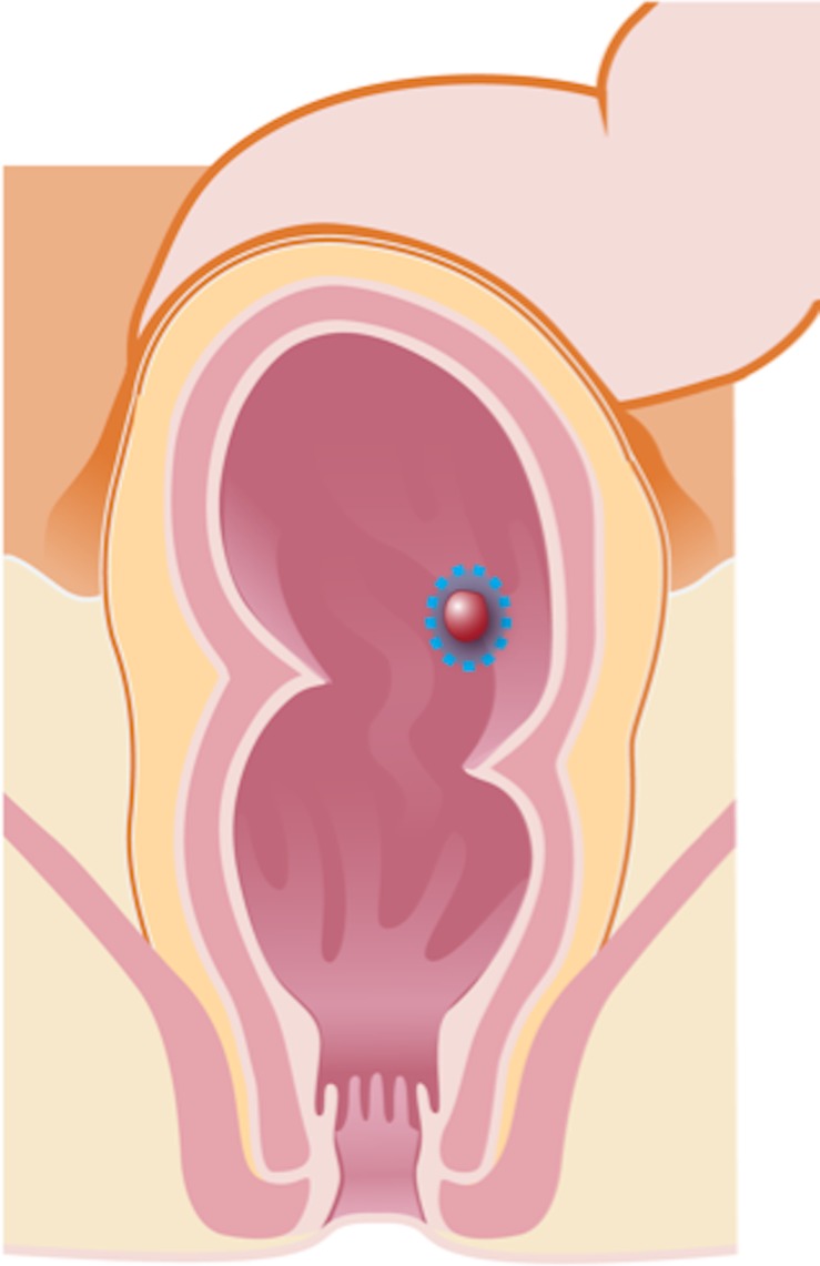
Figure 2a.

Illustrations of the anatomy of the rectum depict various surgical techniques used to treat rectal cancer. Dotted blue lines = anatomic structures removed during the procedure. Red area = rectal tumor. (a) Illustration shows a transanal endoscopic microsurgery with focal endoscopic resection of a tumor. (b) Illustration depicts a low anterior resection and TME and resection of the whole sigmoid or part of it, which preserves the sphincter complex. (c) Illustration depicts an abdominoperineal resection and TME, with resection of the sphincter complex. (d) Illustration depicts an intersphincteric abdominoperineal resection and TME, with dissection within the intersphincteric plane and a portion of the internal sphincter. The entire external sphincter is preserved. (e) Illustration depicts an extralevator abdominoperineal resection and TME, with a broader dissection of the sphincter complex. (Reprinted, under a CC BY-ND 4.0 license, from Memorial Sloan Kettering Cancer Center.)
