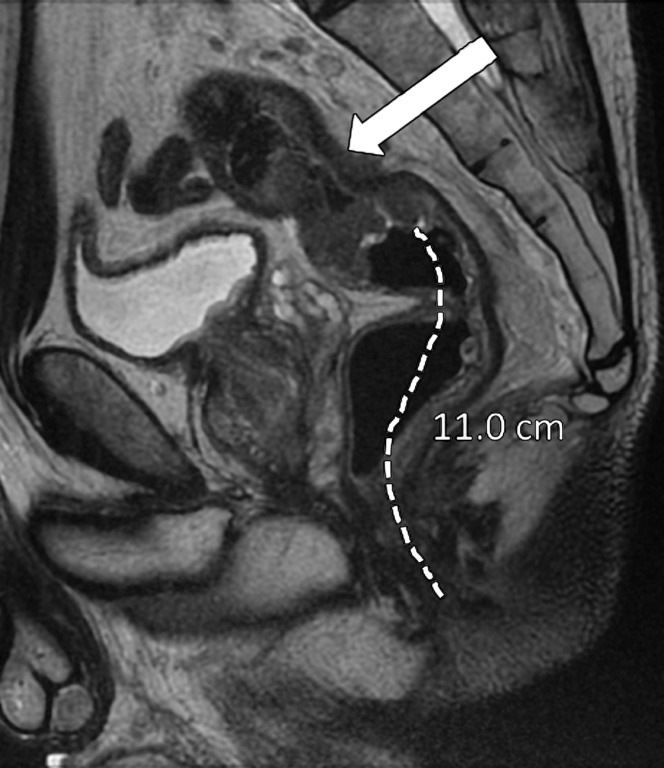
Figure 6b.
Tumor location in the craniocaudal direction. (a) Illustration depicts the sagittal view of the rectum and provides the measurements of the tumor from the anal verge, which help categorize tumor location. Blue lines separate the low, mid-, and high rectum. (Figure 6a reprinted, under a CC BY-ND 4.0 license, from Memorial Sloan Kettering Cancer Center.) (b–d) Sagittal T2-weighted MR images show tumors (arrow) in the high (b), mid- (c), and low (d) rectum. Dotted line = measurement from the rectum entrance to the tumor location.

