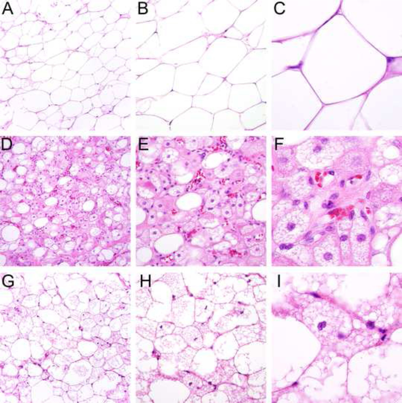
Figure 1.

Morphologic spectrum of hibernoma: tumors consist in variable proportions of mature adipocytes containing a single cytoplasmic vacuole (A-C), large, finely vacuolated cells with eosinophilic granular cytoplasm (hibernoma cells) resembling brown fat (D-F) and multivacuolated lipoblastlike cells with small central nuclei (G-I), which were especially prominent in this case series (see Figure 2).
