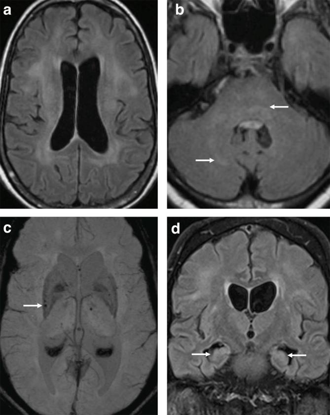
Figure 2.

62-year-old male with West African Trypanosomiasis. (a,b) Axial T2W FLAIR demonstrating ventriculomegaly with bilateral deep white matter hyperintense signal extending from the corona radiata into the posterior limbs of the internal capsules (not shown) as well as the mid brain, pons and right cerebellum (arrows). MRI performed 3 months later shows new susceptibility artefact on SWI in the right posterior putamen (arrow) (c). (d) Coronal T2W FLAIR demonstrates some improvement in bilateral white matter changes and stable ventricular size with hyperintense signal in the hippocampi (arrows). SWI, susceptibility-weighted imaging; T2W FLAIR, T 2 weighted fluid-attenuated inversion recovery.
