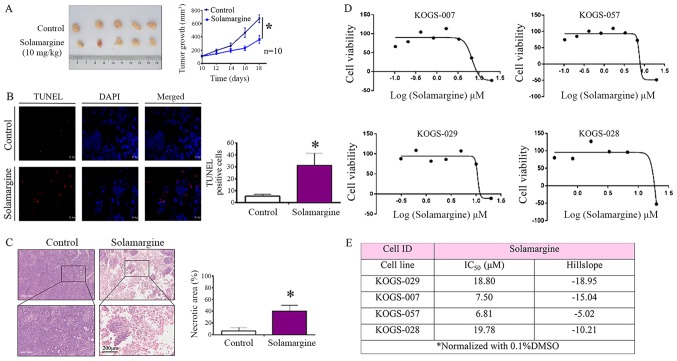
Figure 6.
Effect of Solamargine in a xenograft mouse model and Solamargine sensitivity (IC50) in primary GC cells from patients. (A) Mice were sacrificed and the tumors were collected. Tumor growth was significantly inhibited following treatment with Solamargine. *P<0.05 vs. control. (B) Early apoptosis cells were detected by TUNEL staining. Magnification, ×63. *P<0.05 vs. control. (C) H&E staining was used to analyze the necrotic area (%) of the GC xenograft. Upper panel, magnification, ×5; bottom panel, magnification, ×20. *P<0.05 vs. control. (D) Primary cells from four patients who underwent radical gastrectomy were treated with appropriate doses of Solamargine for 72 h. (E) Solamargine sensitivity (IC50) in primary GC cells from four patients. GC, gastric cancer; IC50, half-maximal inhibitory concentration (48 h); TUNEL, terminal deoxynucleotidyl transferase-mediated dUTP-biotin nick end labeling.

