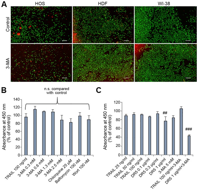
Figure 2.
Normal fibroblasts are resistant to the cytotoxicity of autophagy inhibitors. (A) Images of live/dead assay. HOS cells, human dermal fibroblasts (HDFs) and human WI-38 fibroblasts (WI-38) were treated with 3-MA (2.5 mM) for 24 h at 37°C and stained using a Live/Dead Viability/Cytotoxicity kit. Scale bar, 300 µm. (B and C) HDFs were treated with the agents at the indicated concentrations for 72 h and analyzed for viability using WST-8 assay in triplicate. The data are the means ± SD. Data were analyzed by ANOVA followed by Tukey's post hoc test. ##P<0.01 and ###P<0.001 vs. untreated control (n=3); n.s., not significant. TRAIL, tumor necrosis factor-related apoptosis-inducing ligand; 3-MA, 3-methyladenine; Wort, wortmannin; DR5, death receptor 5.

