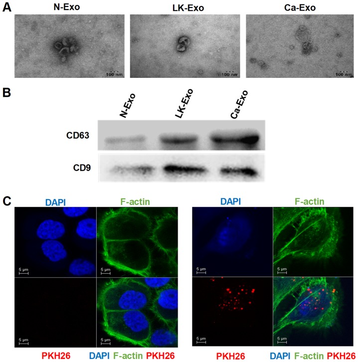
Figure 2.
Identification of exosomes. (A) Exosomes were isolated from MSCs and observed using transmission electron microscopy. (B) The western blotting revealed that the CD63 and CD9 proteins were expressed in the exosomes. (C) Confocal microscopy was used to identify the uptake of PKH26-labeled exosomes, which were secreted by N-MSC and internalized in the cytoplasm. N-exo, exosomes secreted by MSCs derived from normal oral mucosa; LK-exo, exosomes secreted by MSCs derived from oral leukoplakia with dysplasia; Ca-exo, exosomes secreted by MSCs derived from oral carcinoma; MSCs, mesenchymal stem cells; CD63, cluster of differentiation 63.

