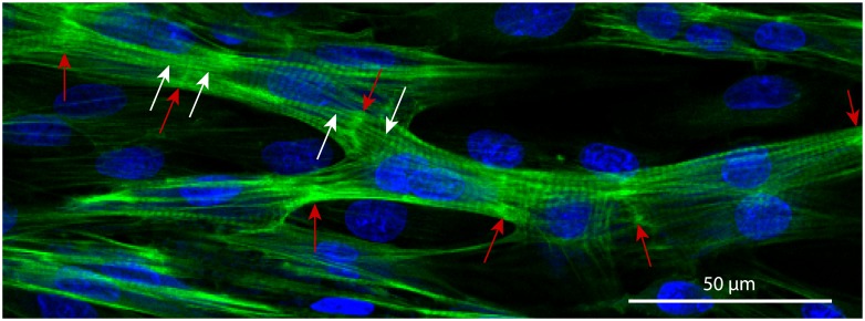
Fig 3. Cardiac syncytium in a 3-days culture of the neonatal rat cardiomyocytes.
The cells have formed intercalated discs (ID, bright-green, highlighted with red arrows), aligned their cytoskeletons (the aligned strains on the both sides from ID are shown with white arrows) and formed a branching network. Nuclei are shown in blue (DAPI, labels DNA), and actin strands are shown in green (phalloidin, labels F-actin).

